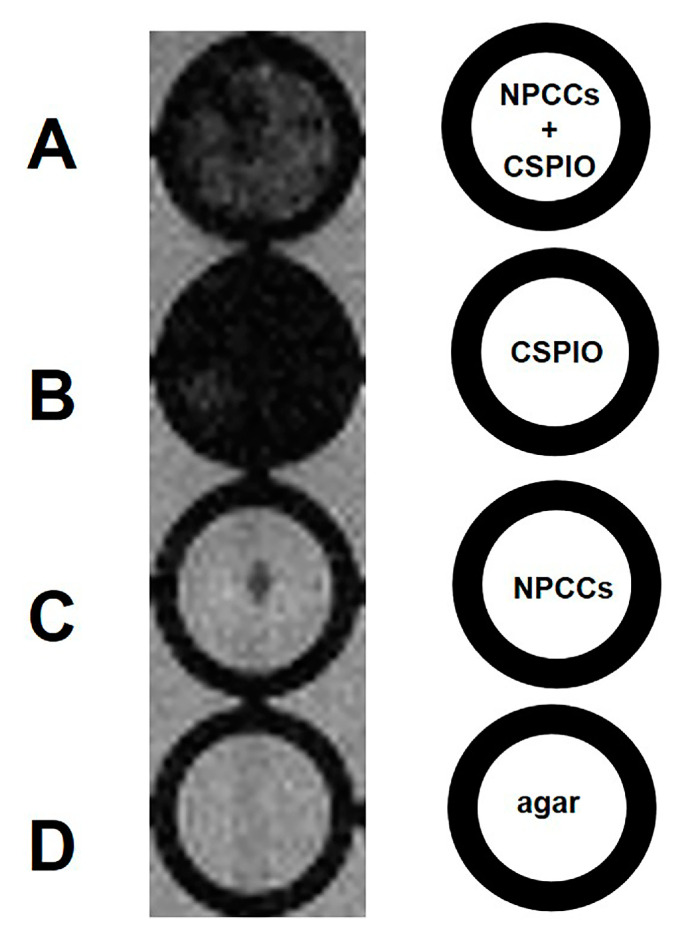
Figure 2.
In vitro MRI of 700 NPCCs incubated overnight with (A) and without (C) chitosan-coated superparamagnetic iron oxide (CSPIO) nanoparticles. CSPIO (B) was used as a positive control and agar (D) was used as a negative control. All were scanned by a 7.0 T MRI system. In contrast to unlabeled NPCCs (C), CSPIO-loaded NPCCs (A) appeared as dark spots.

