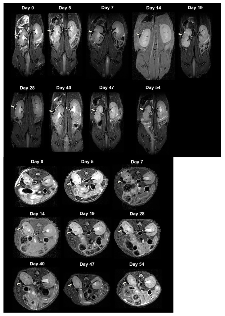
Figure 3.
In vivo MR image of NPCCs after transplantation. Two thousand CSPIO-labeled NPCCs were transplanted under the left kidney capsule of a nude mouse. The recipient was scanned by a 7.0 T MRI system with coronal (upper panel) and transverse (lower panel) sections. The graft of CSPIO-labeled NPCCs was indicated by arrows.

