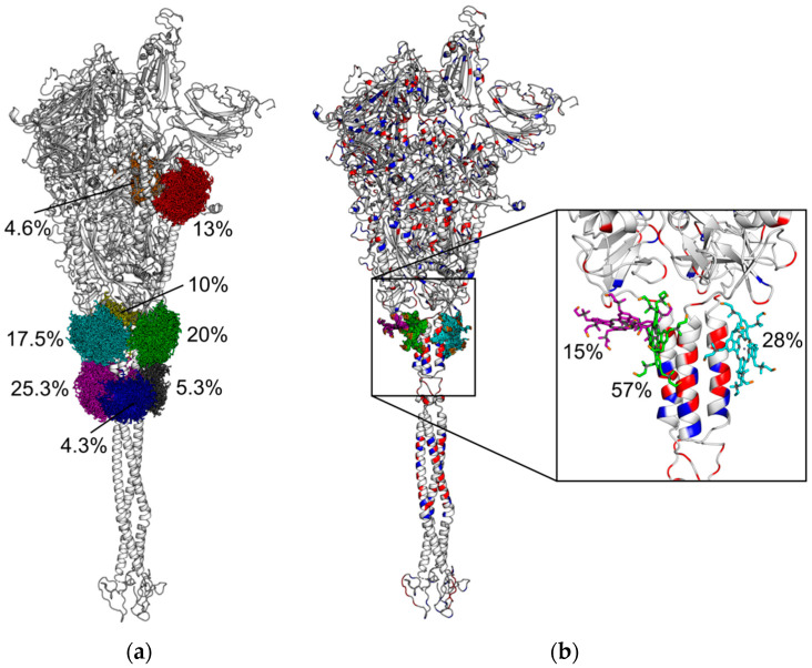
Figure 5.
Cartoon representation of SARS-CoV-2 spike protein with clusters of Zn-PcChol8+ molecules with electrostatic energy of attraction to SARS-CoV-2 spike protein of more than 8 kT (a) and 11 kT (b). The structures of Zn-PcChol8+ molecules belonging to the same cluster are colored by the same color. The relative sizes of clusters, in percentage terms, are given in the figure. The secondary structure of protein in plate (b) is colored by the type of amino acid charge residues (blue are Lys and Arg residues, and red are Glu and Asp residues). The inset shows the magnified area of SARS-CoV-2 spike protein with central structures of three clusters revealed by cluster analysis at attraction electrostatic energy of more than 11 kT.

