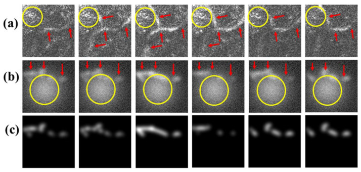
Figure 8.
(a) Six successive en face FF-OCT images of human skin in vivo showing distribution of capillaries with speckle background and (b) six successive images of ACN data showing imitated capillaries with speckle noise in the background; speckle noise is indicated by yellow circle and capillary pathways by red arrows. (c) Corresponding ground truth images of (b).

