Figure 5.
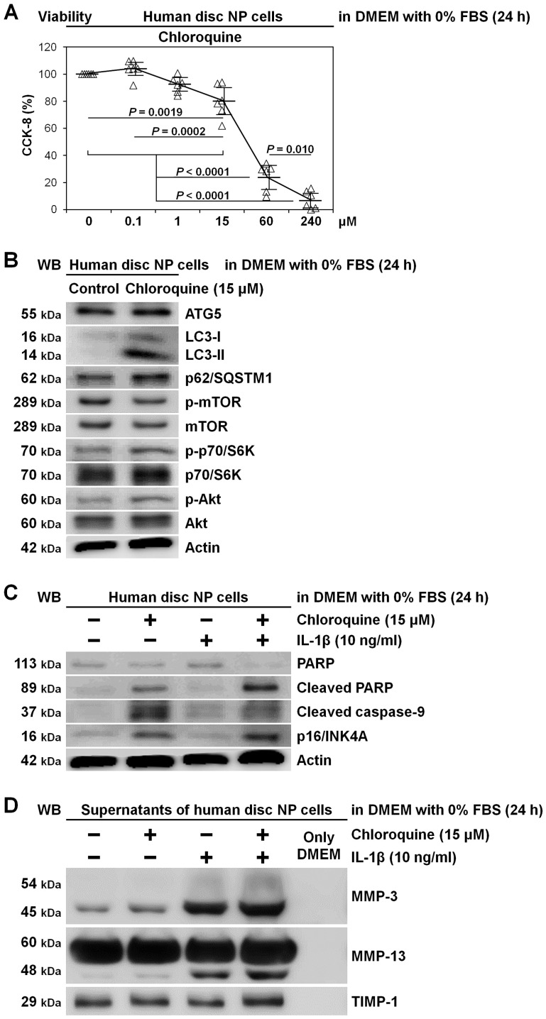
Induced apoptosis and senescence rather than matrix catabolism by autophagy inhibition through chloroquine supplementation in human disc NP cells. (A) Cell viability of human disc NP cells using CCK-8 after treatment of 0–240 μM chloroquine in serum-free DMEM with 0% FBS for 24 h. Changes in CCK-8 dehydrogenase activity of chloroquine treatment relative to the vehicle control are shown. Data are the mean ± 95% CI. One-way repeated-measures ANOVA and the Tukey–Kramer post-hoc test were used (n = 6). (B) Western blotting for autophagic ATG5, LC3, and p62/SQSTM1 and mTOR signaling-related mTOR, phosphorylated mTOR, p70/S6K, phosphorylated p70/S6K, Akt, and phosphorylated Akt in total protein extracts from human disc NP cells after culturing for 24 h in serum-free DMEM with or without 15 μM chloroquine. Actin was used as a loading control. Immunoblots shown are representative of experiments with similar results (n = 6). (C) Western blotting for pro-apoptotic cleaved PARP and cleaved caspase-9, anti-apoptotic PARP, pro-senescent p16/INK4A, and loading control actin in total protein extracts from human disc NP cells after culturing for 24 h in serum-free DMEM with or without 15 μM chloroquine. Immunoblots shown are representative of experiments with similar results (n = 6). (D) Western blotting for catabolic MMP-3 and MMP-13 and anti-catabolic TIMP-1 in supernatant protein extracts from human disc NP cells after 24 h culture in serum-free DMEM with or without 15 μM chloroquine. Immunoblots shown are representative of experiments with similar results (n = 6).

