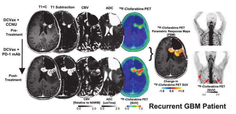
Figure 10.
Post-contrast T1 weighted, T1-subtraction, rCBV, ADC, [18F]-FAC PET + MRI fusion and whole-body maximum intensity projection images of [18F]-CFA from a patient with recurrent GBM before (top) and after (bottom) immunotherapy are shown. Reprinted with permission from ref. [114].

