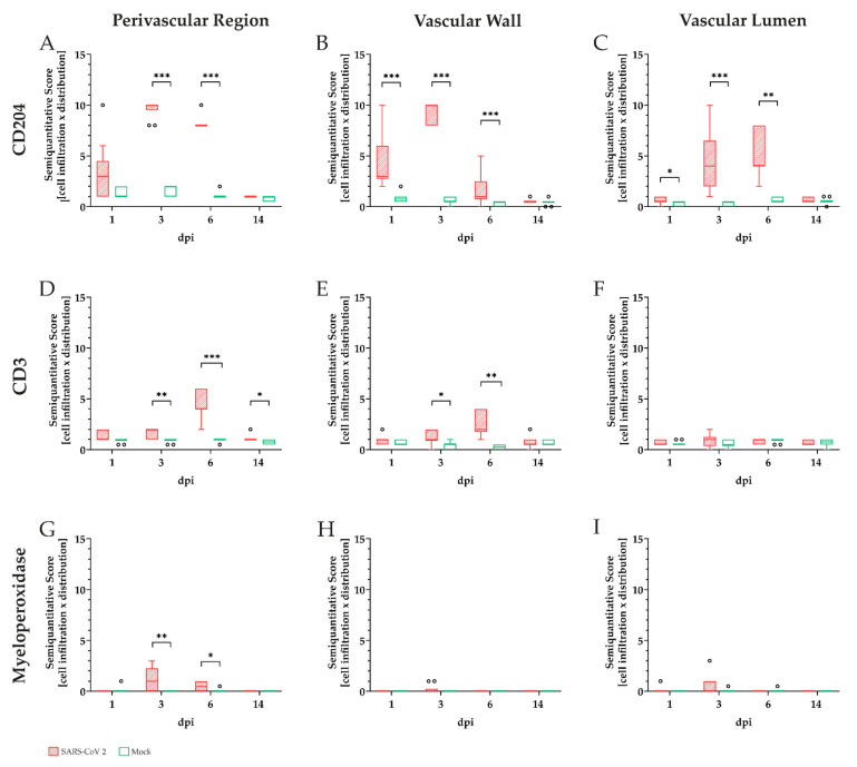
Figure 5.
Semiquantitative analysis of inflammatory cells within different compartments (perivascular, vascular, and intraluminal) in the pulmonary vasculature of golden Syrian hamsters during severe acute respiratory syndrome coronavirus-2 (SARS-CoV-2) in comparison to mock-infected animals. Immunoreactivity of macrophages (CD204, A–C), T-lymphocytes (CD3, D–F), and neutrophils/heterophils (myeloperoxidase, G–I) was examined by light microscopy. Overall immune cell infiltration in respective areas was rated as very mild (0,5), mild (1), mild to moderate (2), moderate (3), mild to severe (4), moderate to severe (5), or severe (6). Signal distribution was assessed as: focal/oligofocal (< 4 foci; (1), multifocal (≥ 4 foci; (2) or diffuse (3). For better comparative interpretation a final score for each area and animal was calculated as the product of cell infiltration and signal distribution. Macrophages (A–C) represented the main infiltrating immune cell population in all analyzed areas, with the highest scores at 3 dpi. Statistically significant infiltration could be observed at 3 and 6 dpi in the perivascular region (A) and at 1, 3, and 6 dpi in the vascular wall (B). Perivascular and vascular detection of T-lymphocytes (D,E) was overall lower at all time points compared to macrophages, with significant infiltration detectable at 3, 6, and 14 dpi in the perivascular region (D) and at 3 and 6 dpi in the vascular wall (E). Significant levels of neutrophils/heterophils were mainly located perivascularly and detected at 3 and 6 dpi (G). Data are shown as box and whisker plots with mean and quartiles. Significant differences between the groups obtained by the Kruskal–Wallis test followed by Dunn-Bonferroni post hoc testing are indicated by * (* p ≤ 0.05, ** p ≤ 0.01, *** p ≤ 0.001).

