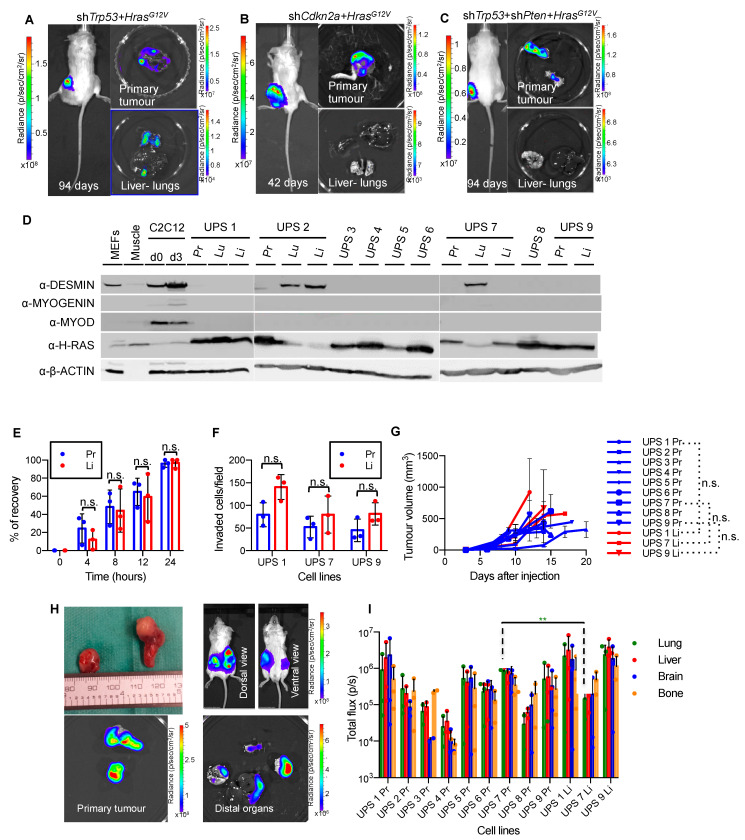
Figure 1.
Establishing an HrasG12V-driven autochthonous metastatic UPS model. (A–C). Representative bioluminescence imaging of living mice and of primary tumors, lungs and livers of tumor-bearing SCID/Beige mice induced by intramuscular injection of MuLE viruses expressing luciferase plus either shRNA-Trp53 + HrasG12V (A) shRNA-Cdkn2a + HrasG12V (B) or shRNA-Trp53 + shRNA-Pten + HrasG12V (C). (D). Protein immunoblotting for the indicated antibodies on cells isolated from primary UPS tumors (Pr) and metastatic lesions from lungs (Lu) and livers (Li), as well as on mouse embryo fibroblasts (MEFs), mouse muscle and C2C12 myoblast cells at day 0 and day 3 of myogenic differentiation as controls. VINCULIN is used as loading control. (E). Time course of recovery of wounds induced by scratch assay on confluent cells. Experiments were performed on UPS 7 Pr and UPS 7 Li. Results are representative of similar experiments performed using UPS 1 Pr and UPS 1 Li and using UPS 9 Pr and UPS 9 Li (Figure S2). Mean ± std. dev. are shown (n = 3). In all matching pairs of cells, the differences between primary tumor and metastatic cell lines were not statistically significant (p > 0.05, unpaired t-test Holm-Sidak method). (F). Overnight trans-well migration assay. Data is derived from analyses of ten independent fields of 1.5 mm2 per membrane and is depicted as mean ± std. dev. of 3 biological replicates. Differences were not statistically significant (p > 0.05, two-way ANOVA, Sidak method). (G). Growth curves of primary tumors arising from subcutaneous injection of the indicated primary and metastatic cell lines. Differences between growth rates of tumors arising from metastatic lesions over their corresponding primary tumor cell lines were not statistically significant (n.s., Welch’s t-test). (H). Example of primary tumor and metastatic lesions arising within 20 days after subcutaneous injection of cells isolated from Trp53 + HrasG12V primary tumor. Distal organs include lungs, liver, brain and femur (bone marrow). (I). Luciferase intensity from metastatic lesions arising from subcutaneous allografts of primary tumor or metastatic lesion cells. Mean ± std. dev. of each organ from 3 injected mice per cell line are shown. Luciferase intensity in lung arising from cell line UPS 7 Pr was significantly higher than the luciferase intensity in the lung from its metastatic counterpart (UPS 7 Li) (** p < 0.01, unpaired t-test Holm-Sidak method). All other comparisons were not statistically significant.

