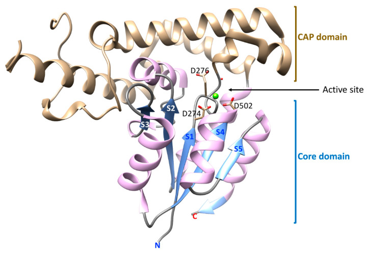
Figure 3.
A ribbon drawing of the EYA2 ED domain generated using UCSF Chimera [17] from 3GEB.PDB [15]. The Mg2+ ion is depicted as a green sphere. The core and cap domains, as discussed here, are shown. β-strands that make up the Rossmanoid HAD core structure are numbered S1–S5, going from N- to C-terminus of the domain. Amino acid side-chains in the active site (the nucleophilic Asp and the genral acid-base Asp in Motif I, and the Asp in Motif IV that co-ordinates the metal ion) are shown in the active site.

