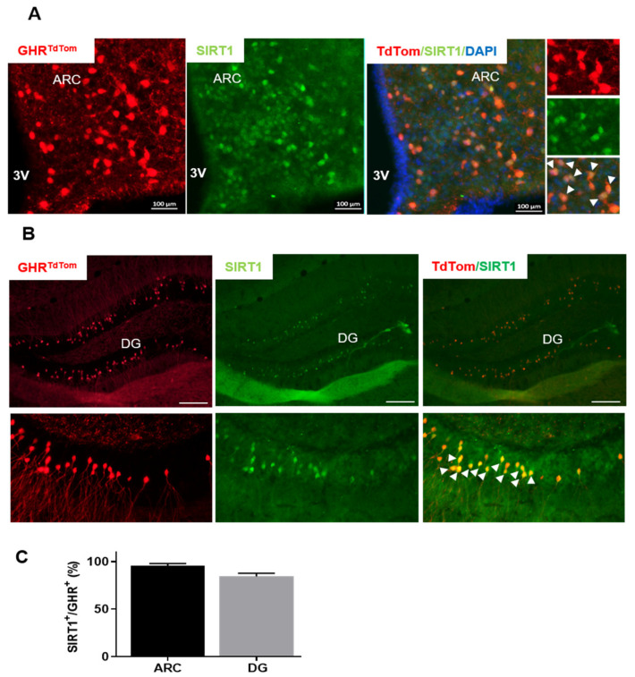
Figure 1.
Sirtuin 1 (SIRT1) is co-localized with growth hormone receptor (GHR)+ neurons. Representative fluorescence microscopy images of (A) the hypothalamus and (B) the hippocampus of fasted GHRtdTom mice. Red, tdTomato; green, SIRT1; the merged image shows co-localization between GHR and SIRT1 in the arcuate nucleus (ARC). The magnified images on the right side show the co-localization of tdTomato and SIRT1 fluorescence in the GHR+ neurons (arrows). DG, dentate gyrus; ARH or ARC, arcuate nucleus of the hypothalamus. (C) The percentage of the GHRSIRT+ subpopulation from the total GHR+ neurons in the ARC or DG. The data are shown as mean ± SEM. n = 4–5. The data were analyzed from both male and female mice. Scale bar: 100 µm. The 3V, third ventricle.

