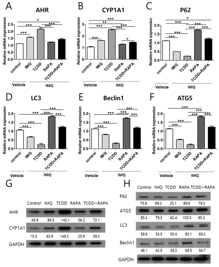
Figure 5.
mRNA and Western blot analysis of AHR, CYP1A1, and autophagy-related factor expression changes in skin tissue with treatment with vehicle control, IMQ, IMQ + TCDD, and IMQ + Rapa, or IMQ + TCDD + Rapa. RNA and protein were extracted from the back skin. (A–F) qPCR and (G,H) Western blot analysis of AHR, CYP1A1, P62, ATG5, LC3, and Beclin1 expressions in mice treated with 5 different regimens (n = 5 per treatment group). qPCR data represent the mean ± standard deviation (SD) of three independent experiments (each performed in duplicate). Statistical differences were determined by one-way analysis of variance (ANOVA) followed by post hoc Dunnett’s test. * p < 0.05; ** p < 0.01; *** p < 0.001. Western blot normalization was based on GAPDH. Western blot data are representative of three independent experiments. IMQ: imiquimod, Rapa: rapamycin.

