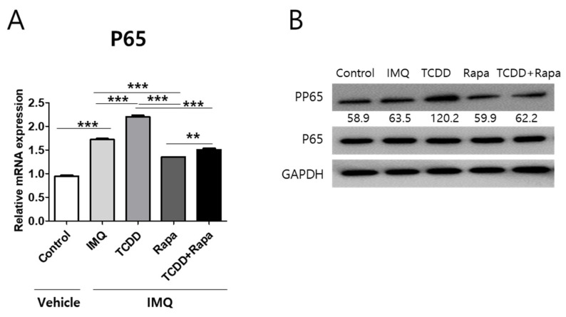
Figure 9.
The effects of TCDD, rapamycin, or a combination of both on P65 mRNA and phosphorylation of P65 expression changes in mouse skin tissue. (A) qPCR of P65 and (B) Western blot analysis of phosphorylation of P65 expression in mouse skin from 5 different treatment regimens (n = 5 per group). Data represent the mean ± SD of three independent experiments (each performed in duplicate). Statistical differences were determined by one-way analysis of variance (ANOVA) followed by post hoc Dunnett’s test. ** p < 0.01; *** p < 0.001. Western blot normalization was based on GAPDH. Western blot data are representative of three independent experiments. IMQ: imiquimod, Rapa: rapamycin.

