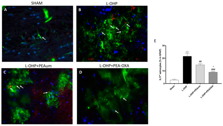
Figure 5.
Effect of PEA-OXA on GFAP and IL-17 expression. The immunofluorescence staining for GFAP (green) and IL-17 (red) was made in the lumbar spinal cord sections. Considerable astrogliosis with production of IL17 was present in L-OHP panels (B,E), compared to the sham group (A,E). The treatment with PEA-OXA did not show any significant immunoreactivity of IL-17 in astrocytes (D,E), much more then PEAum (C,E). Scale bar = 20 μm (particle). Data are means ± SEM from n = 10 mice/group. Counting of colocalized cells confirmed our data (E). The colocalization image was analyzed with image J software. *** p < 0.001 vs. sham; ## p < 0.01 and ### p < 0.001 vs. L-OHP; ^ p < 0.05 vs. PEAum.

