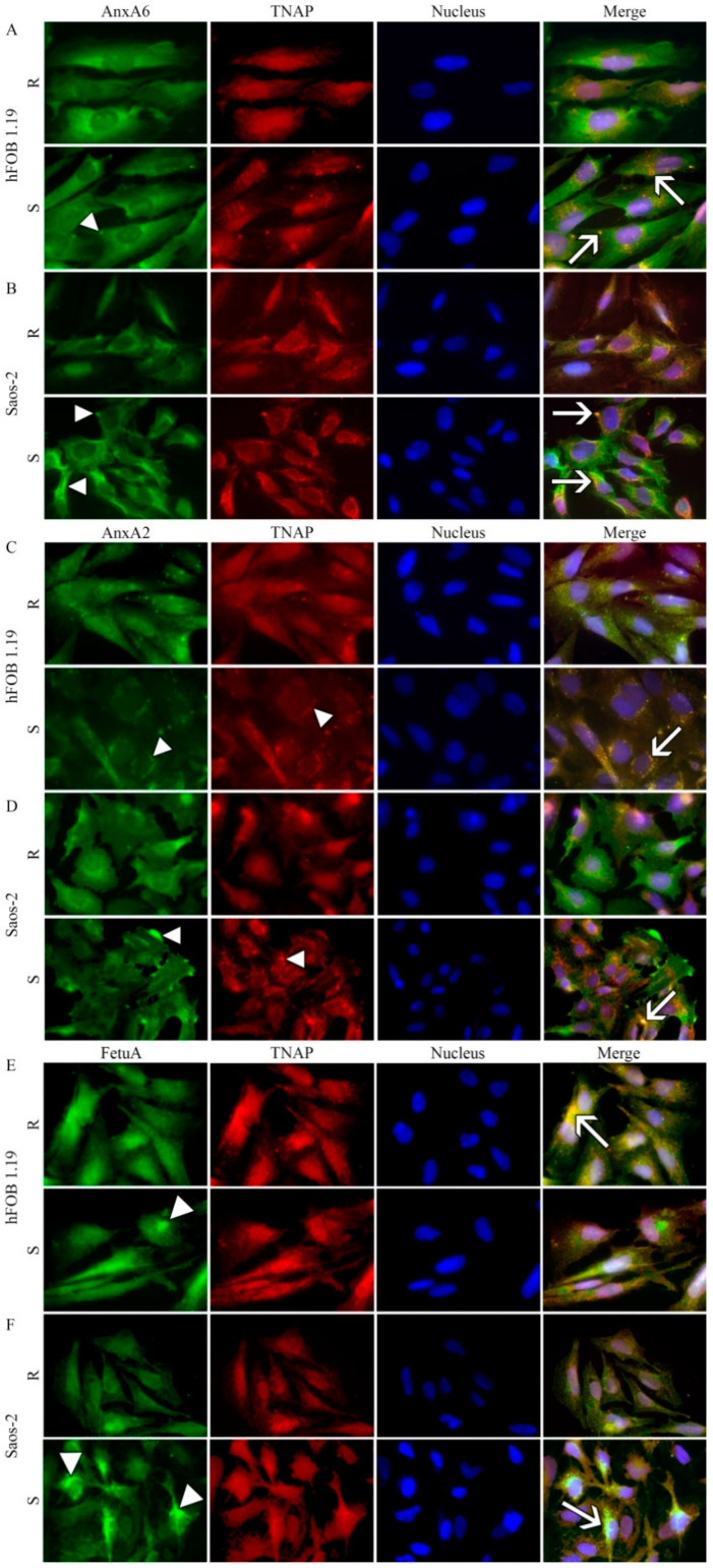
Figure 4.
Co-localization of AnxA6, AnxA2, or fetuin-A (FetuA) with TNAP in hFOB 1.19 (A,C,E) and Saos-2 (B,D,F) cells in resting conditions (R) or after seven-day stimulation with AA and β-GP (S). The cells were incubated with appropriate antibodies: mouse monoclonal anti-AnxA6 (A,B), mouse monoclonal anti-AnxA2 (C,D), or mouse monoclonal anti-FetuA (E,F), all followed by goat anti-mouse IgG-FITC (green); rabbit polyclonal anti-TNAP followed by goat anti-rabbit IgG-TRITC (red) and DAPI for nuclei (blue) and observed under an Axio Observer.Z1 FM (Carl Zeiss, Poznan, Poland) with Phase contrast and appropriate fluorescent filters, magnification 630 x. Arrowheads indicate protein accumulation in vesicular and/or cluster structures. The yellow color and arrows on the merge images indicate AnxA6 (A,B), AnxA2 (C,D), or FetuA (E,F) co-localization with TNAP. Results of a typical experiment are presented.

