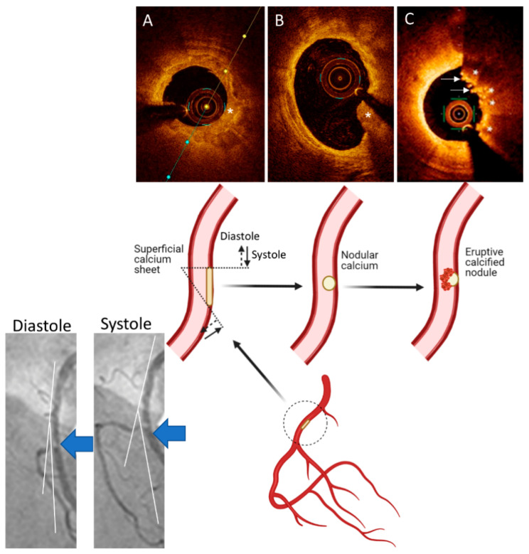
Figure 3.
A Working Hypothesis for Formation of Eruptive Calcified Nodules. Superficial calcified sheets (asterisk) (A) located in the arterial segments with hinge movement, e.g., in the mid segment of the right coronary artery (large blue arrows on the angiographic pictures and inset), are subject to cyclic mechanical forces during systole and diastole, which could weaken and fragment the sheets of calcium, thus resulting in protruding nodular calcium (asterisk) (B) that is surrounded by fibrin. These nodules eventually erupt through the plaque surface ((C), asterisks), causing disruption in the intima, with superimposition of thrombus ((C), arrows). Parts of the figure are adapted from Lee et al. [55] with permission.

