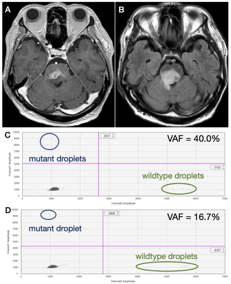
Figure 3.
A recurrent DMG case in which H3F3A K27M was suspected. MR images show (A) a heterogeneously enhancing lesion with possible necrosis and (B) perifocal hyperintensity on fluid attenuated inversion recovery (FLAIR). Since mutant droplets were detected from both wells by ddPCR analysis, H3F3A K27M mutation was strongly considered. However, since only (C) two and (D) one mutant droplets were detected from each well, a definitive diagnosis was not achieved.

