Figure 2.
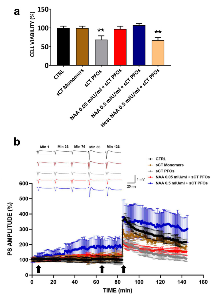
(a) Cell viability assessment in HT22 cells treated with Neuraminidase (NAA) and salmon Calcitonin (sCT): HT22 cells pre-treated with NAA at concentrations of 0.05 mIU/mL (red bar, n = 21 from N = 5 experiments) and 0.5 mIU/mL (blue bar, n = 16 from N = 4 experiments) for 1 h and then incubated with Prefibrillar Oligomers of salmon Calcitonin (sCT PFOs) for 24 h showed cell viability comparable to that of the CTRL group (black bar, n = 34 from N = 5 experiments). Treatment with sCT PFOs (grey bar, n = 20 from N = 5 experiments) significantly reduced cell survival by approximately 30% (** p < 0.01), whereas treatment with sCT monomers (brown bar, n = 17 from N = 4 experiments) did not affect cell viability. Finally, pre-treatment with heat NAA 0.5 mIU/mL + sCT PFOs (pink bar, n = 14 from N = 3 experiments) induced a reduction in cell viability of approximately 30%, like that obtained when HT22 cells were treated with sCT PFOs (** p < 0.01). (b) Synaptic plasticity in CA1 subfield of mouse hippocampal slices following NAA and sCT administration: % Population Spike (PS) amplitude as a function of time after tetanic stimulation (HFS), applied at time 𝑡 = 86 min (arrow), following NAA (𝑡 = 5 min, arrow) and sCT (𝑡 = 65 min, arrow) administration, is shown in CTRL (black bar, n = 6), in sCT monomers (brown bar, n = 4), in sCT PFOs (grey bar, n = 4), in NAA 0.05 mIU/mL + sCT PFOs (red bar, n = 7), and in NAA 0.5 mIU/mL + sCT PFOs (blue bar, n = 5) mice slices at minutes 1, 36, 76, 86 and 136. The insert shows representative recordings obtained from slices of each experimental group; curves of each group refer to PS at times 1, 36, 76, 86 and 136 min.

