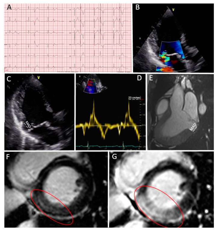
Figure 2.
Case 1: Primary prevention ICD implantation after multi-modality imaging work-up. (A) Patient’s ECG; (B) moderate mitral regurgitation during color Doppler transthoracic echocardiography; (C) bileaflet prolapse with MAD (in white); (D) “Pickelhaube sign” during transthoracic echocardiography; (E) MAD measured in a steady-state free precession three-chamber view during CMR; (F,G) a large zone of fibrosis in the basal inferior wall in LGE sequences (red circle).

