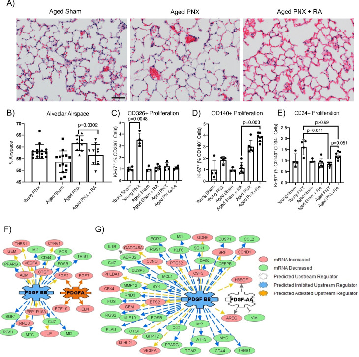
Figure 1.
Mouse lung regeneration following RA pretreatment. (A) H&E stain of aged Sham, aged mice and RA-pretreated mice 21 days after PNX. Scale bar is 200 µm. (B) Quantification of alveolar space by morphometric point and intersection counting analysis (PNX21). (C–E) Proliferation was assessed by KI67 expression by flow cytometry (PNX5). Data were normalised to respective sham controls. (C) CD326+, (D) CD140+ and (E) CD140+CD34+ cells in young and aged Sham, PNX and RA pretreatment mice. (F, G) Upstream analyses of RNA sequencing gene expression changes in PDGFRA+ mesenchymal cells from ‘aged mice’ when compared with either ‘young mice’ or ‘RA pretreatment’ mice (PNX5). In both comparisons, genes downstream of PDGF signalling were significantly altered. (F) In PDGFRA+ mesenchymal cells isolated from RA pretreatment, PDGF-BB signalling was predicted to be significantly inhibited, while PDGFA signalling was activated. (G) Similarly, in young mice compared with aged mice, PDGF-BB signalling was predicted to be significantly inhibited, with PDGFA signalling being altered in PDGFRA+ mesenchymal cells. mRNA, messenger RNA; PDGF, platelet-derived growth factor; PDGFA, platelet-derived growth factor subunit A; PNX, partial pneumonectomy; RA, retinoic acid.

