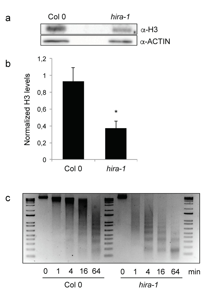
Figure 2.
Loss of HIRA leads to reduced H3 content and MNase hypersensitivity. (a) Representative Western blot of H3 histones in total protein extracts from WT and hira-1 mutant dry seeds. (b) Quantification of H3 band intensities relative to ACTIN from three biological and four technical replicates. One wild type sample was set to 1 in each technical replicate. Statistically significant differences relative to WT were determined using a two-sided Student’s t-test (* p < 0.05). (c) Nuclei from WT or hira-1 mutant seeds were isolated and incubated with MNase for 1, 4, 16 and 64 min. Equal amounts of digested DNA were loaded on an agarose gel and stained with ethidium bromide. One experiment of three biological replicates with similar results is shown.

