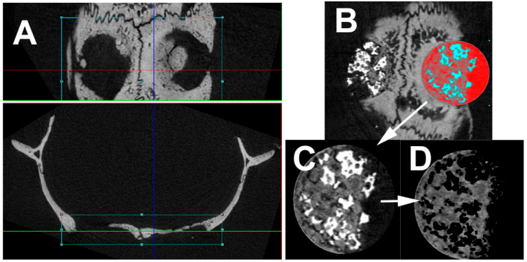
Figure 2.
Picture showing the micro-computed tomography (CT) evaluation. (A) First the slices have been placed in correct orientation for reconstruction. (B) Thereafter a circular region of interest (diameter 5 mm) has been placed into the defect. (C) by defining the first and last slice with bone, the volume of interest was defined. (D) Semi-automated evaluation protocol removed very dense regions (Actifuse, NanoBone®) as well as air and evaluated the volume of interest with respect to the defined parameters.

