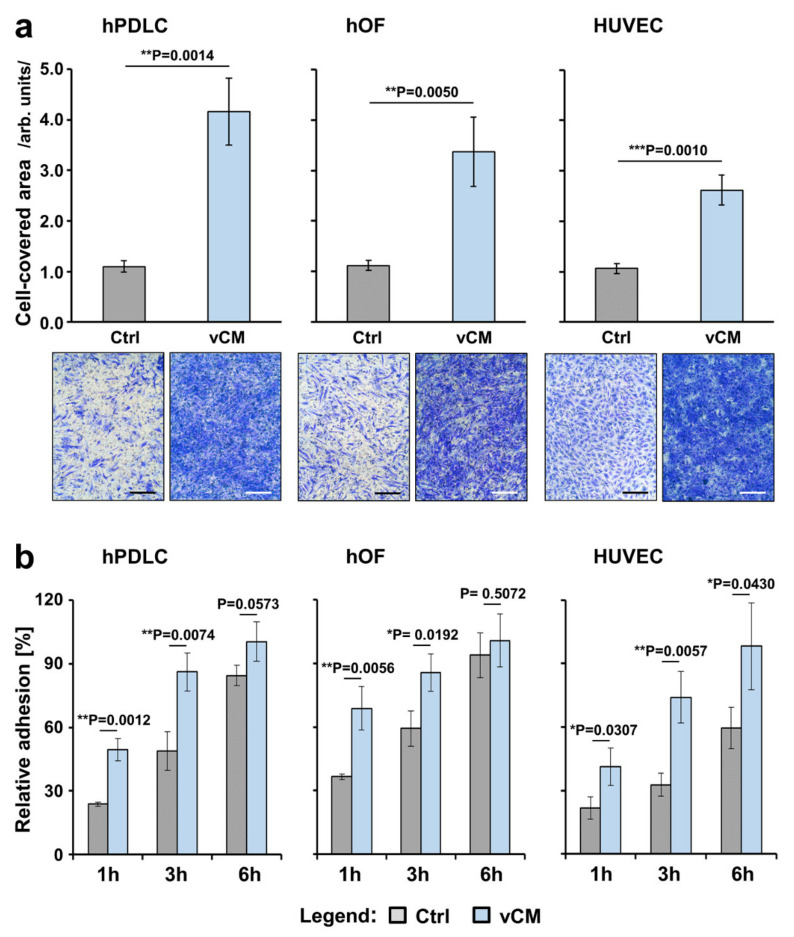
Figure 1.
Promigratory (a) and proadhesive (b) effects of vCM on three primary cell types of human origin. (a) Migratory capacity of hPDLCs (left panel), hOFs (middle panel), and HUVECs (right panel) toward vCM was examined by modified Boyden chamber assay using cell culture inserts with permeable membranes (8 µm pore size). Following an incubation period of 18 h, cells that had migrated through the membrane were stained and quantified by using ImageJ. Cells that migrated in the absence of vCM served as a control (Ctrl). Means ± SD from three independent experiments performed in duplicates and a significant difference between the control and test group, ** p < 0.01, *** p < 0.001 are shown; Representative images of crystal violet-stained cells in each of the experimental groups are shown below the bar charts. Scale bar, 500 μm. (b) Adhesive capacity of hPDLCs (left panel), hOFs (middle panel), and HUVECs (right panel) on vCM was examined using crystal violet staining of adherent cells at 1, 3, and 6 h postseeding. Controls (Ctrl) represent cells of each cell type seeded on tissue culture-treated plates in the absence of vCM for the indicated times. Normalization of the experimental values to the values obtained for the total number of seeded control cells (taken as 100% adhesion) was applied. Means ± SD from three independent experiments performed in duplicates and significant differences to control cells at each time point, ** p < 0.01, * p < 0.05 are shown.

