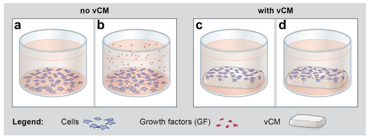
Figure 3.
Schematic representation of the conditions under which hPDLCs, hOFs, and HUVECs were seeded for subsequent qRT-PCR analyses of the anti- and pro- fibrinolytic gene expression presented in Figure 4, Figure 5, and Figure 6. (a) Starved cells were grown for 48 h on tissue culture plastic in the absence of vCM and growth factors (GF), a condition further denoted as a control (Ctrl). (b) Starved cells were grown for 48 h on tissue culture plastic in the presence of GF applied in suspension. (c) Starved cells were grown for 48 h on native/uncoated vCM placed in ultra-low attachment plates. (d) Starved cells were grown for 48 h on vCM coated with EMD or each of the four recombinant growth factors, TGF-β1, PDGF-BB, FGF-2, or GDF-5 in ultra-low attachment plates. Please note that the scheme illustrates the conditions upon cell seeding and therefore, released from the vCM growth factors are not depicted in the cell culture medium in (d). In addition, there is no obvious difference in the appearance between tissue culture-treated plates for adhesive culture (a,b) and ultra-low attachment plates (c,d).

