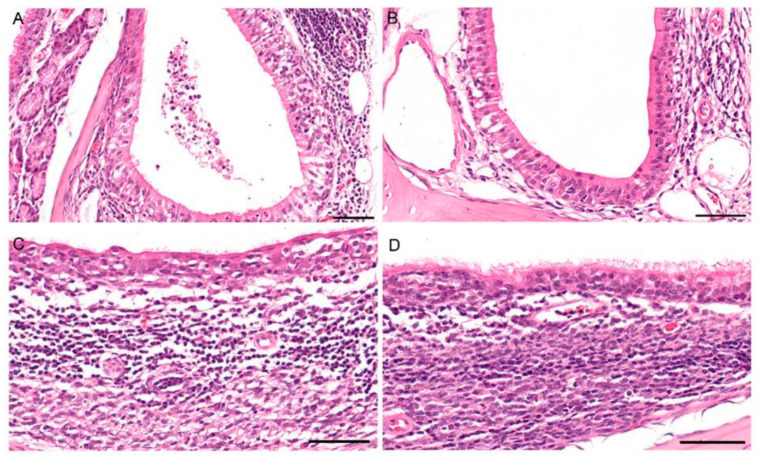
Figure 7.
Histopathology in the nasal mucosa of fruit bats after infection with bat-origin H9N2 (A/bat/Egypt/381OP/2017), 7 dpi. (A) Inoculated animal #5: moderate transmigration of granulocytes and lymphocytes as well as intraluminal cellular debris within the nasolacrimal duct. (B) Unaffected nasolacrimal duct for comparison. (C) Contact animal #6: loss of cilia and moderate degeneration of the mucosal epithelium of the nasal-associated lymphoid tissue (NALT). (D) Unaffected NALT for comparison. All bars = 50 µm.

