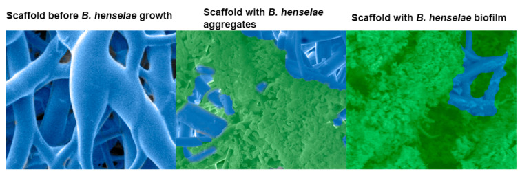
Figure 1.
Scanning electron micrograph of a 48 h Bartonella henselae (B. henselae) biofilm growing on a 3-dimensional nanofibrous scaffold. Left, scaffold before bacterial growth. Middle, bacterial growth, adhesion, and aggregation around the scaffold branches. Right, B. henselae biofilm covering the scaffold and eclipsing the bacterial cells. Biofilm was preserved by the addition of the cationic dye, Alcian blue.

