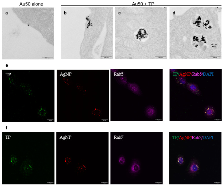
Figure 4.
Imaging of endocytic structures for bystander uptake. CHO cells were incubated with Au50 alone (a) or Au50 + TP (b–d) in the serum-free DMEM medium for 2 h before fixation and being processed for TEM imaging. Ultrastructurally, AuNPs were found in macropinosome-like endocytic vacuoles (>200 nm in diameter) (b), early endosomes (c) and late endosomal multivesicular bodies (d). Presentative images were shown here. Scale bars, 500 nm. (e,f) Colocalization of bystander AgNP with endosome markers as revealed by confocal imaging. Hela cells were incubated with FAM-TP (green) and bystander AgNP (AgNP-555, red) for 1 h. Endosome markers (magenta) were stained by immunofluorescence, as described in the Methods. Bystander AgNPs were caputured in Rab5-localized early endosomes (e) and Rab7-localized late endosomes (f). Scale bars, 20 μm.

