
Figure 7.

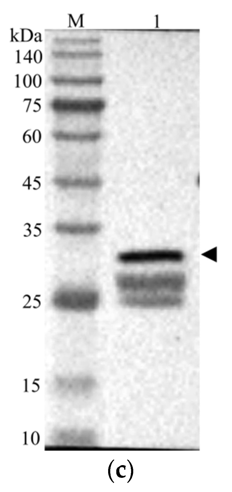
Identification of recombinant CcLys2 protein. (a) SDS-PAGE analysis of the bacterial lysate. Lane 1: the non-induced bacterial culture; Lane 2: the soluble supernatant; Lanes 3 and 4: inclusion body protein. (b) Purified CcLys2. Lane 1: fusion CcLys2. (c) Western blot analysis of CcLys2. Lane 1: blotting band of fusion CcLys2. M: Protein molecular weight marker (10–140 kDa). The arrow indicates the bands of fusion CcLys2.