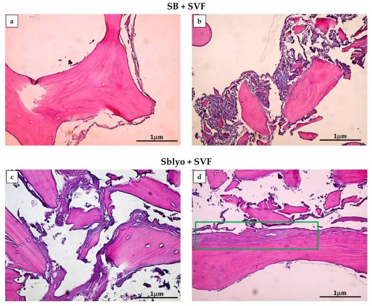
Figure 5.
H&E staining. New tissue formation is evident on both SB (a,b) and SBlyo (c,d) after 60 days of culture. Panels (a,b) show cell growth in the periphery and among bone trabeculae, respectively. On SBlyo (d), a well-organized tissue portion is visible. The green rectangle indicates an area with well-organized neo-formed tissue. Magnification 20×. Scale bar: 1 µm.

