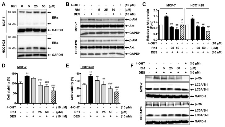
Figure 6.
Rh1 showed a competitive effect with an estrogenic agent on BC cells. (A) MCF-7 cells and HCC1428 cells were treated with different concentrations of Rh1 (5, 25, and 50 µM) for 24 h. Total cell lysates then analyzed the estrogen receptor alpha (Erα) protein expression using Western blotting. (B,C) MCF-7 cells and HCC1428 cells were treated with 10-nM DES in the presence or absence of Rh1 (5, 25, and 50 µM) or 10-µM 4-OHT for 30 min. Total cell lysates were then analyzed using Western blotting (B). The relative quantification of protein levels (C) was analyzed using Image J software. (D,E) MCF-7 cells (D) and HCC1428 cells (E) were treated with 10-nM DES in the presence or absence of Rh1 (5, 25, and 50 µM) or 10-µM 4-OHT for 24 h. The 3-(4, 5-dimethylthiazole-2-yl)-2, 5-diphenyltetrazolium bromide (MTT) assay was used to analyze the cell viability. (F) MCF-7 cells and HCC1428 cells were treated with 10-nM DES in the presence or absence of Rh1 (5, 25, and 50 µM) or 10-µM 4-OHT for 12 h. Total cell lysates were then analyzed using Western blotting. (C–E) Data are presented as the mean ± SD (n = 3), * p < 0.05 and ** p < 0.01 compared with the control sample and # p < 0.05, ## p < 0.01 and ### p < 0.001 compared with the DES-treated sample, respectively. 4-OHT: 4-hydroxytamoxifen and DES: diethylstilbestrol.

