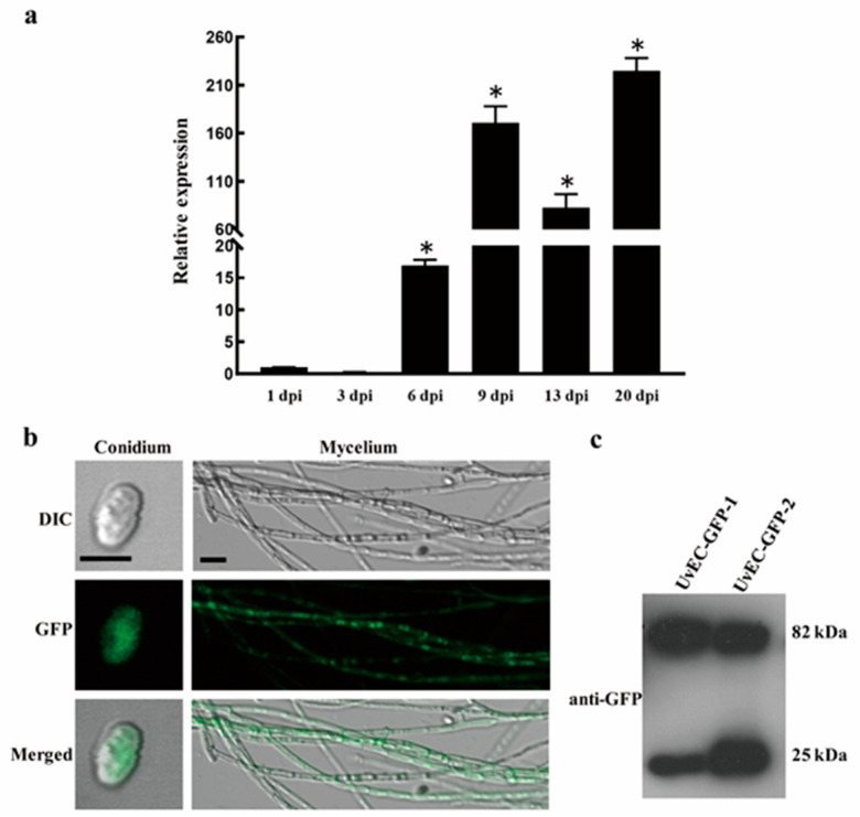
Figure 6.
Expression of UvEC1 and subcellular localization of UvEC1 in U. virens. (a) Relative expression of UvEC1 at different infection stages on rice spikelets (1–21 d), as determined by RT-qPCR. UvEC1 expression was normalized to that of β-tubulin. Asterisks represent significant differences relative to 1 dpi by LSD at p = 0.05 (b) Subcellular localization of UvEC1 in U. virens. DIC, differential interference contrast; GFP, green fluorescent protein. Scale bars = 10 μm. (c) Immunoblot analysis of UvEC1-GFP-CDC11 in the C∆UvEC1-1 strain.

