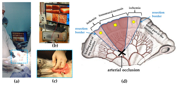
Figure 1.
The procedure for intraoperative small intestine visualization using multimodal optical coherence tomography (MM OCT). (a) General overview of the trans-serous MM OCT study; (b) MM OCT system (1) with probe (2) and an example of data set acquired after tissue scanning (3); (c) the probe (2) in a sterile sheath is placed on the surface of the normal ileum; (d) diagram of the MM OCT study of ischemic bowel: square mark—point of the OCT probe localization on the anesthetized part of the intestine, round marks—points from which MM OCT images were obtained from the ischemic areas of the intestine to be resected.

