Figure 2.
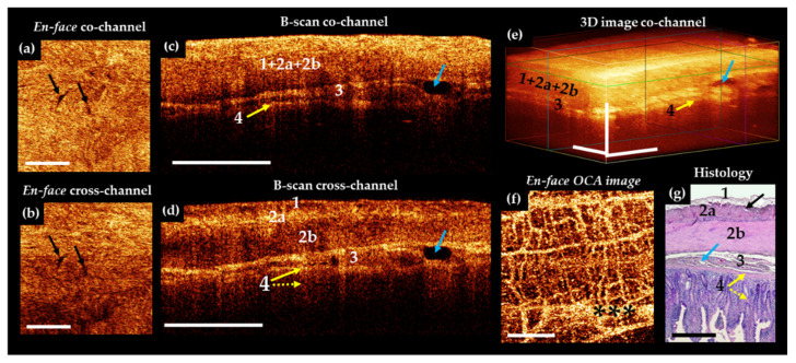
Representative trans-serosal MM OCT images (a–f) and corresponding histology (g) of normal small intestine of a pig. In the B-scans cross—channel (d) all 4 layers—serous membrane (1), muscularis externa (longitudinal muscular layer 2a and circular muscular layer 2b), submucosa (3) with blood and lymphatic vessels (blue arrows) and mucosa (4) (in particular, muscularis mucosa (yellow solid arrow) and adjacent to it the lamina propria (yellow dotted arrow)) are more clearly resolved, while in the co-channel not all anatomical layers are optically separated (c). (a,b) En-face OCT images at the level of the longitudinal muscular layer (green horizontal line in (e)). Intermuscular spaces look like slits and there are few of them (black arrows). In the OCTA image (f), the network of blood vessels is dense and branched. Large and small blood vessels are visualized; the background of the image is light due to a dense network of a functioning capillary network. (g) Histological specimens stained with H&E. Asterisks in (f) indicate artefact of motion. Scale bars in CP OCT and OCTA images 1 mm, in histology 0.5 mm.

