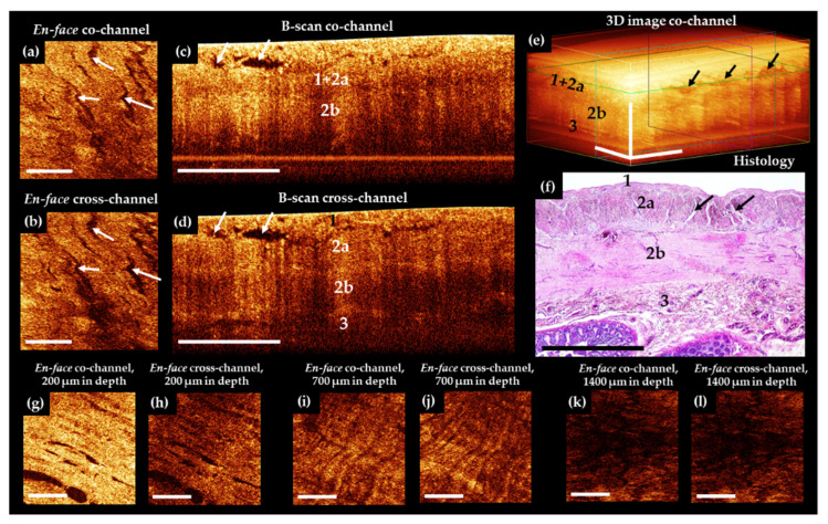
Figure 3.
Representative trans-serosal CP OCT images of normal human ileum. In the B-scan cross-channel (d) the serous membrane (1), muscularis externa (longitudinal muscular layer 2a and circular muscular layer 2b) and the boundary with submucosa (3) are more clearly resolved, while in the co-channel, not all anatomical layers are optically separated (c,e). (a,b) En-face OCT images at the level of the longitudinal muscular layer (green horizontal line in (e)). Intermuscular spaces ((f), black arrows) look like slits in CP OCT images ((a–d), white arrows; (e), black arrows) and they are located within the outer muscular layer. (f) Corresponding histological image, H&E staining. In some cases, a clear change in the direction of muscle bundles in different layers (g–j) and a characteristic pattern of the lamina propria (k,l) immediately under the muscularis mucosa can be observed. Scale bars 1 mm.

