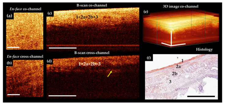
Figure 5.
Representative trans-serosal CP OCT images of ileum with transmural ischemic necrosis. In the B-scans co– (c) and cross– (d) channels and in 3D OCT image (e) the depth of tissue visualization is decreased significantly and individual layers of the serous membrane (1), outer longitudinal muscular layer (2a), and inner circular muscular layer (2b) are not distinguishable due to the developed necrosis in all layers of the intestinal wall (f). In the B-scans cross-channel (d) in the lower part of the inner muscle layer, the signal is noticeably reduced, and the transition from this layer to underlying tissue of the submucosa (3) can be detected. (a,b) En-face OCT images at the level of the longitudinal muscle layer (green horizontal line in (e)), demarcated from the surrounding tissue fluid accumulation is not observed. (f) Corresponding histological image, H&E staining. Scale bars 1 mm.

