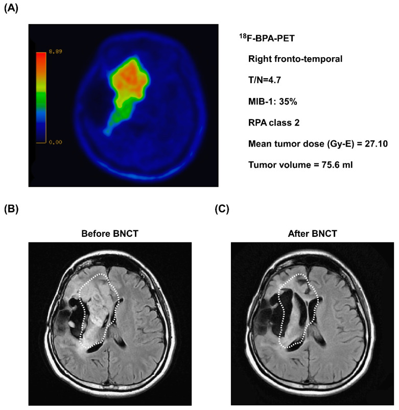
Figure 5.
Representative images of tumor shrinkage after BNCT treatment. (A) Before BNCT treatment, 18F-BPA-PET revealed the location of strong tumor activity in the right fronto-temporal lobe. (B) MRI imaging of the patient before BNCT treatment. An anaplastic astrocytoma was observed in the right temporal lobe (white dashed circles). (C) Three months after BNCT treatment, a follow-up MRI image of the patient showed a significant reduction in tumor volume (white dashed circles).

