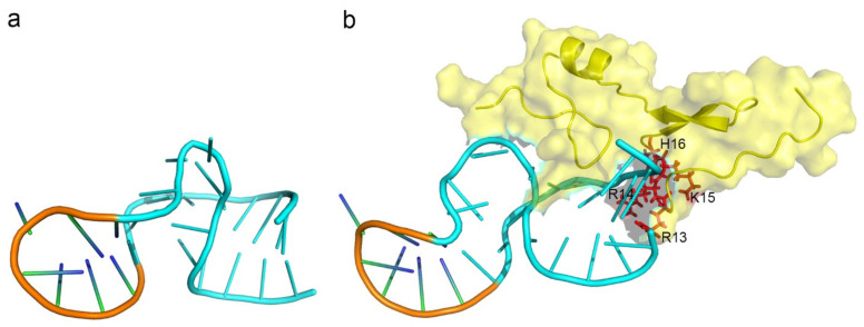
Figure 4.
The model structure of Macugen (pegaptanib) and the induced fit-binding interaction with its target protein. (a) The structure model of the aptamer, pegaptanib, was shown according to the secondary structure calculated by NMR [131]. The conformation was the free form of the aptamer. (b) The interface of the aptamer in complex with the target protein, VEGF165 (PDB ID: 2VGH). The structure and the surface of VEGF165 was shown in yellow. The residues on pegaptanib that undergo a conformational change are shown in blue, while the other residues were shown in orange. The residues on VEGF165 that participated in the interaction of the complex were shown in red and labelled in black (R13, R14, K15, H16).

