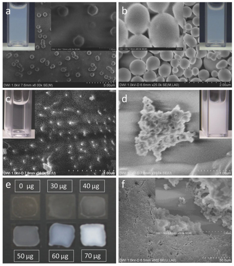
Figure 5.
Field-emission-scanning electron microscope (FESEM) images of 0.24 mmol β-CD-MA (DS2) nanogels before (a) and after complexation with 70 μg mL−1 chlorhexidine (CHX) on aluminum surface (b). Cryo-FESEM image of 0.47 mmol β -CD-MA (DS4) nanogels before (c) and after complexation with 70 μg mL−1 CHX (d). The inset in (a–d) shows a dispersion of the β-CD-MA nanogels in a cuvette. Photography of 0.47 mmol β-CD-MA (DS4) nanogels with different CHX content coated on glass plates (e) and FESEM images of the nanogel film consisting of the 0.47 mmol CD-MA (DS4) nanogels with 70 μg mL−1 CHX (f). The second insets in (a,b,f) show enlarged images of the nanogels. Reproduced with permission from [111], Macromolecular Bioscience, 2017.

