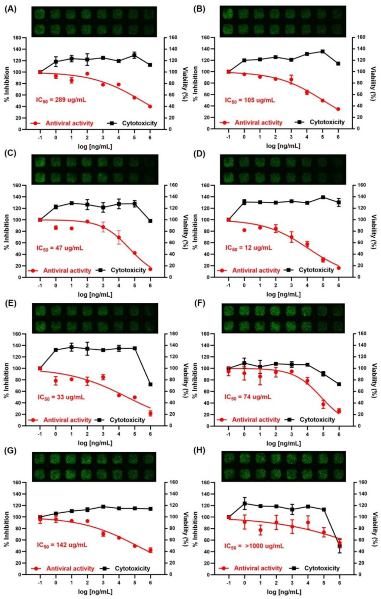
Figure 2.
Determination of the cytotoxicity (right y axis, black squares) and antiviral activity (left y axis, red circles) of the crude polysaccharides in HEK293/ACE2 cells. The viability of HEK293/ACE2 cells was assessed using an CellTiter-Glo® Luminescent cell viability assay kit (Promega, Madison, WI, USA) after treatment with the indicated concentrations of the crude polysaccharides from (A) Undaria pinnatifida sporophyll, (B) Laminaria japonica, (C) Hizikia fusiforme, (D) Sargassum horneri, (E) abalone viscera, (F) Codium fragile, (G) fucoidan (Herim), and (H) Porphyra tenera for 96 h. The inhibition of viral infection by the crude polysaccharides was performed with a SARS-CoV-2 pseudovirus (COV-PS02). Results are expressed as a percent of inhibition in drug-treated cultures versus untreated and were plotted with Graphpad prism software (Graph-Pad, San Diego, CA, USA). Values are the means ± S.D. (n = 3). The viral infection of HEK293/ACE2 cells was detected as a GFP fluorescence by an MBD ASFA scanner (MBD Biotech., Swon, South Korea) and was presented on the top of each graph.

