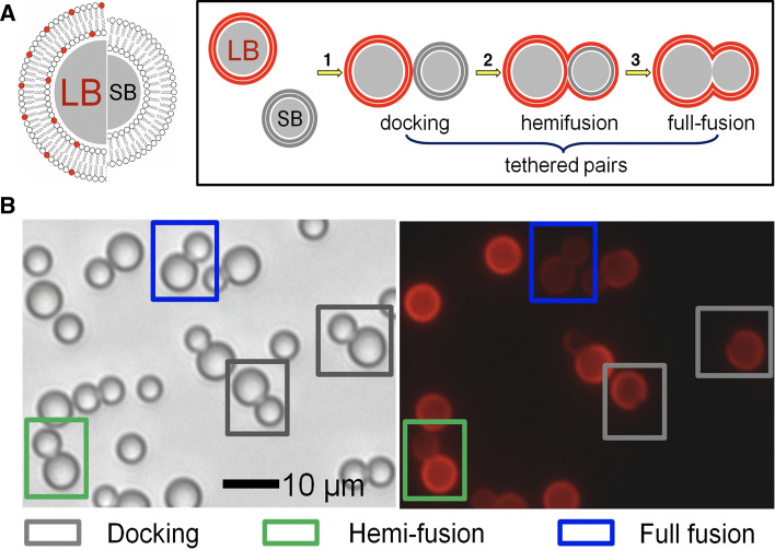
Fig. 8.
a Illustration of large and small silica beads coated with a lipid bilayer (LB, diameter 6.5 m and SB, diameter 4.7 m). Only the large bead (LB) is coated with a fluorescently labeled membrane. The distinct intermediate states such as docking (1), hemifusion (2), and full fusion (2) can be distinguished unequivocally by a combination of bright-field and fluorescence microscopy. b Two micrographs showing a bright-field (left) image and from the same spot also a fluorescence image (right) of LB-i-K3 and SB-i-E3 on a glassy surface. Docked pairs (gray box), hemifused pairs (green box), as well as fully fused pairs (blue box) are discernible (from Bao et al. (2013))

