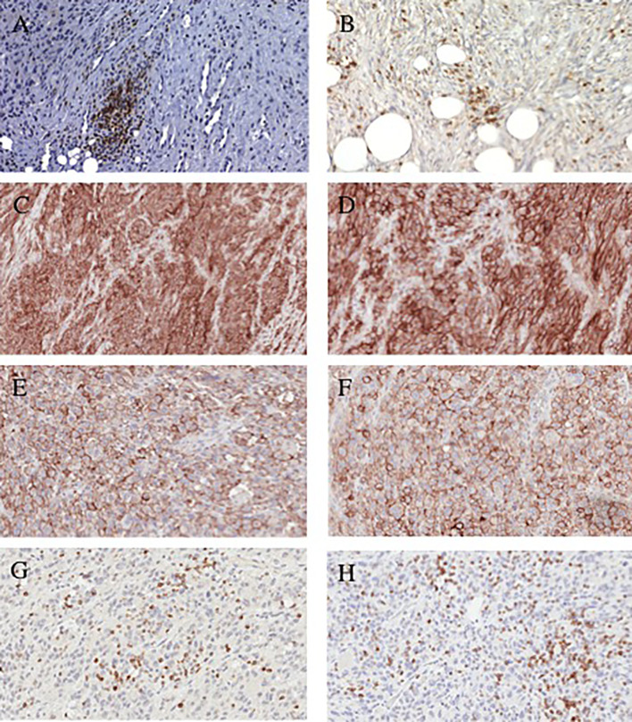
Figure 1.
Representative immonohistochemical staining patterns of dedifferentiated chondrosarcoma tumors with lymphocyte and checkpoint-specific monoclonal antibodies. (A) CD4+ TILs (200x magnification). (B) CD8+ TILs (200x magnification). (C) PD-L1 positive cells (100x magnification). (D) PD-L1 positive cells (200x magnification). (E) PD-L1 positive cells (200x magnification). (F); PD-L1 positive cells (200x magnification). (G) PD-1 positive lymphocytes (200x magnification). (H) PD-1 positive lymphocytes (200x magnification).

