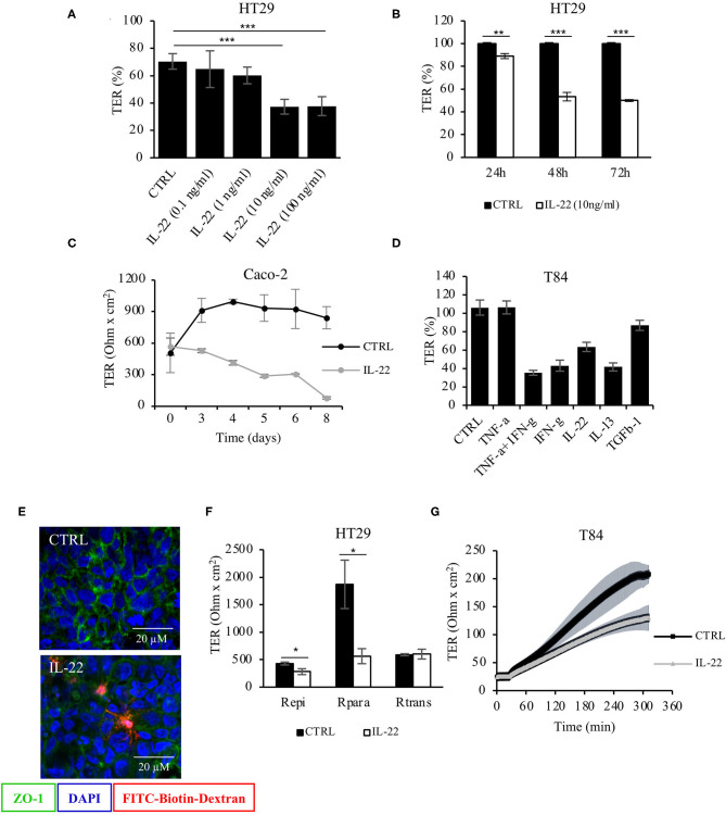
Figure 1.
Barrier integrity is affected by IL-22. (A) Transepithelial resistance (TER) was determined in HT-29/B6 cells grown on transwell filters. Cells were exposed to IL-22 at different concentrations (0.1, 1, 10, and 100 ng/ml). TERs after 72 h of IL-22 exposure are shown n = 25. Mann–Whitney U test; ***p < 0.001. (B) TER time course in HT29/B6 cells exposed to IL-22 (10 ng/ml); n = 32. Mann–Whitney U test; **p < 0.01; ***p < 0.001. (C) TER measured in CaCo-2 cells exposed to IL-22 (10 ng/ml) for a longer time course (up to 8 days); n = 3. (D) Comparative analysis of TER in T84 cells (grown on transwell filters) after a 48 h-exposure to various cytokines (TNFα: 1,000 U/ml, IFNγ: 100 U/ml, IL-22: 10 ng/ml; IL-13: 10 ng/ml; TGF-b1: 20 ng/ml); n = 8. (E) Sandwich assay revealing transepithelial passage of macromolecules, specifically TexasRed-dextran3000 (red fluorescence) in control and IL-22-treated CaCo-2 cells. E-cadherin, green; nuclei, blue; n = 3. (F) Two-path impedance analysis: HT-29/B6 cells grown on transwell filters were exposed to IL-22 (10 ng/ml) for 48 h. After mounting filters to Ussing chambers paracellular and transcellular components of TER were determined by two-path impedance; n = 6. Mann Whitney U test; *p < 0.05 (G) Calcium switch experiment: T84 cells growing on transwell filters were exposed to IL-22 (10 ng/ml, 48 h) and mounted to Ussing chambers, where TER was monitored in 10 s-intervals throughout the experiment. Transepithelial resistance was measured every 60 min for 6 h; n = 3. Mann–Whitney U-test; *p < 0.05; **p < 0.01; ***p < 0.001.

