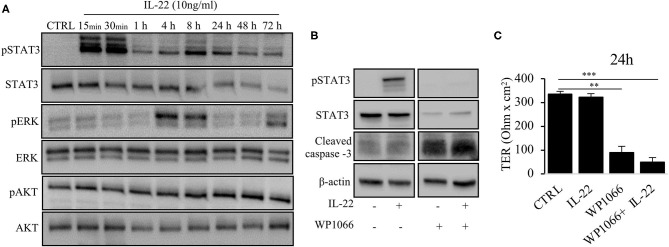
Figure 6.
IL-22 induces STAT3 and ERK phosphorylation. (A) HT-29/B6 cells were exposed to IL-22 (10 ng/ml) for the indicated times. Subsequently, cells were lysed and protein levels of STAT3, ERK and AKT and their phospho-levels were investigated by Western blotting. Representative blots of three independent experiments are shown. (B) HT-29/B6 cells were incubated in the presence of the STAT3 inhibitor WP1066 (50 μM) for 2 h and IL-22 (10 ng/ml) for 1 h as indicated. Western blotting of cell lysates was performed to quantify protein levels of STAT3 total, phospho-STAT3 (pSTAT3), cleaved caspase-3, and b-actin as loading control. (C) TER was determined after 24 h of IL-22 exposure of HT-29/B6 cells growing on transwell filters treated with IL-22 (10 ng/ml), WP1066 (50 μM) as indicated. Mann–Whitney U test; n = 3; *p < 0.05; **p < 0.01; ***p < 0.001.

