Fig. 3.
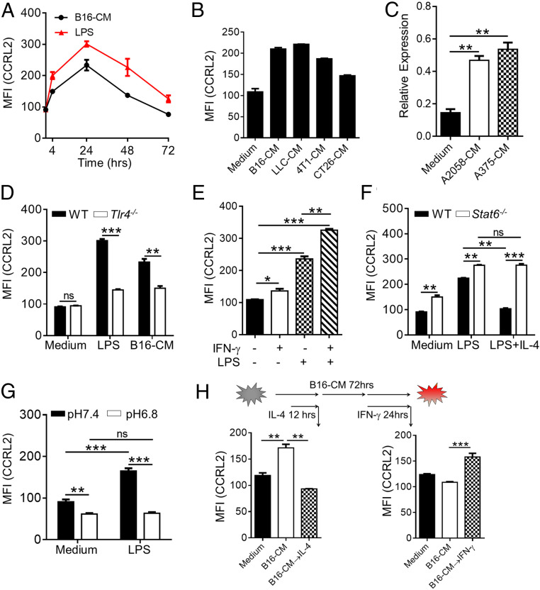
The immunostimulatory factors induce CCRL2 expression in macrophages, which is antagonized by immunosuppressive factors. (A and B) CCRL2 MFI in WT bone marrow-derived macrophages (BMDM) stimulated with LPS (100 ng/mL) or condition media (CM) of B16F10 cells (B16-CM) (A), and in WT BMDM with CM from different tumor cell lines for 24 h (B). (C) Quantitative polymerase chain reaction (qPCR) analysis of CCRL2 expression in THP1-derived human macrophages stimulated with CM of human melanoma cell lines. (D–G) CCRL2 MFI expression in WT and Tlr4−/− BMDM following stimulation with LPS or B16-CM (D), in WT BMDM with LPS, IFN-γ or LPS, and IFN-γ together (E), in WT and Stat6−/− BMDM with LPS alone or LPS and IL-4 (20 ng/mL) together (F), and in WT BMDM with LPS (in medium with pH 7.4 or pH 6.8) (G). (H) CCRL2 MFI in WT BMDM that were stimulated with B16-CM for 12 h or 48 h, and then treated with/without IL-4 for another 12 h or with/without IFN-γ for another 24 h along with B16-CM. Data represent mean ± SEM of triplicate wells from a representative of three independent experiments. *P < 0.05; **P < 0.01; ***P < 0.001; ns, not significant.

