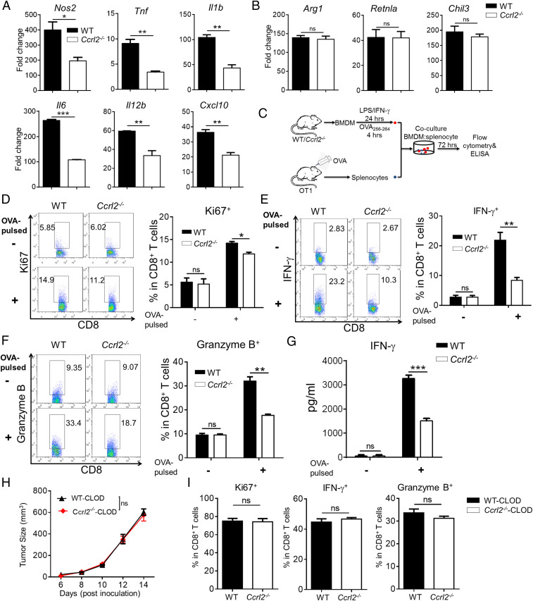
Fig. 6.
CCRL2 promotes activation of M1 macrophages to potentiate anti-tumor CD8+ T-cell responses. (A and B) qPCR analysis of M1-related genes (A) and M2-related genes (B) in WT and Ccrl2−/− BMDM stimulated with LPS/IFN-γ and IL-4, respectively. (C) Schematic protocol for coculture of WT or Ccrl2− BMDM and OT1 splenocytes. (D–F) CD8+ T cells were gated in OT1 splenocytes that were cocultured with LPS/IFN-γ-activated WT or Ccrl2−/− BMDM without or with SIINFEKL (OVA257–264) pulse and examined for expression of Ki67 (D), IFN-γ (E), and Granzyme B (F). (G) The concentrations of IFN-γ in the cocultures determined by ELISA. Data represent mean ± SEM of triplicate wells from a representative of two independent experiments. (H–I) Tumor growth curve (H) and expression of Ki67, IFN-γ, and Granzyme B in CD8+ T cells (I) from clodronate liposomes (CLOD)-treated WT or Ccrl2−/− mice on day 14. In A and B, data represent mean ± SEM of triplicate wells from a representative of three independent experiments. In D–I, data represent mean ± SEM (n = 5 to 6) and similar results were obtained from two to three independent experiments. *P < 0.05; **P < 0.01; ***P < 0.001; ns, not significant.

