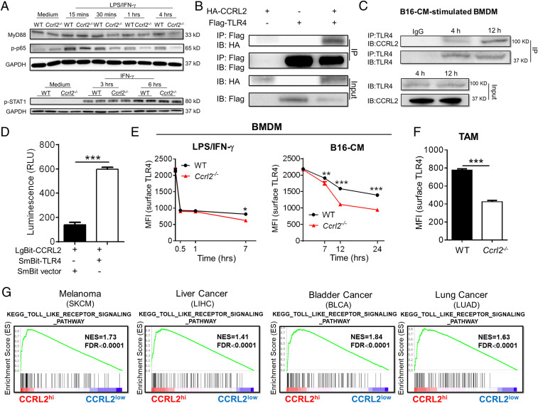
Fig. 7.
CCRL2 retains membreane TLR4 expression to amplify downstream Myd88-NF-κB signaling. (A) Representative Western blots showing protein levels of Myd88, phosphorylated p65 (p-p65), and phosphorylated STAT1 (p-STAT1) in WT and Ccrl2−/− BMDM. (B and C) Coimmunoprecipitation (co-IP) assay showing the interaction between TLR4 and CCRL2 in HEK 293T cells (B) and in BMDM stimulated with B16-CM for indicated time (C). (D) NanoBit proximity assay system showing the interaction between TLR4 and CCRL2 in living cells. Luminescence was assayed using Nano-Glo Live cell substrate. (E and F) Surface TLR4 levels in WT and Ccrl2−/− BMDM (E), and in macrophages from WT and Ccrl2−/− tumors on day 14. Data represent mean ± SEM, n = 4 (F). (G) GSEA demonstrating the enrichment of signature genes of TLR signaling pathway in CCRL2hi tumors. In A–C, representative data from two to three experiments are shown. In D and E, data represent mean ± SEM of triplicate wells from a representative of three independent experiments. **P < 0.01; ***P < 0.001.

