Fig. 3.
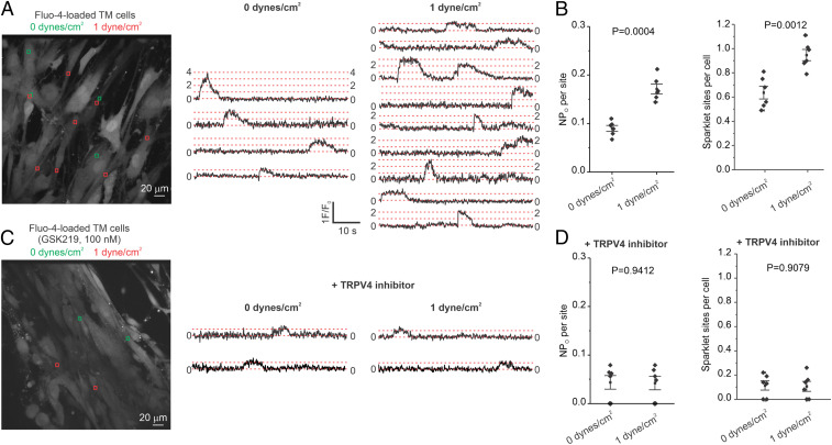
Flow/shear stress increases TRPV4 channel activity in TM cells. (A, Left) A grayscale image of fluo-4–loaded TM cells; the ROIs represent TRPV4 sparklet sites in the absence of flow (green square boxes; 0 dyne/cm2) or the presence of flow/shear stress (red square boxes; 1 dyne/cm2); (A, Right) F/F0 traces from the ROIs shown in the grayscale image indicate TRPV4 sparklet activity before (0 dyne/cm2) and after (1 dyne/cm2) flow/shear stress. (B) Averaged TRPV4 sparklet activity (Left) or the number of sparklet sites per cell (Right) in the absence (0 dyne/cm2, n = 6) or presence of flow (1 dyne/cm2, n = 6). (C, Left) A grayscale image of fluo-4–loaded TM cells in the presence of GSK219 (TRPV4 inhibitor, 100 nM); the ROIs represent TRPV4 sparklet sites in the absence (green square boxes; 0 dyne/cm2) and or presence of flow/shear stress (red square boxes; 1 dyne/cm2). (C, Right) F/F0 traces from the ROIs on the grayscale image indicate TRPV4 sparklet activity before (0 dyne/cm2) and after (1 dyne/cm2) flow/shear stress. Experiments were performed in the presence of the TRPV4 channel inhibitor GSK219 (100 nM). (D) Averaged TRPV4 sparklet activity (Left) or the number of sparklet sites per cell (Right) in the absence (0 dyne/cm2, n = 6) or presence of flow (1 dyne/cm2, n = 6). Experiments were performed in the presence of the TRPV4 channel inhibitor GSK219. Data are presented as mean ± SEM.

