Fig. 8.
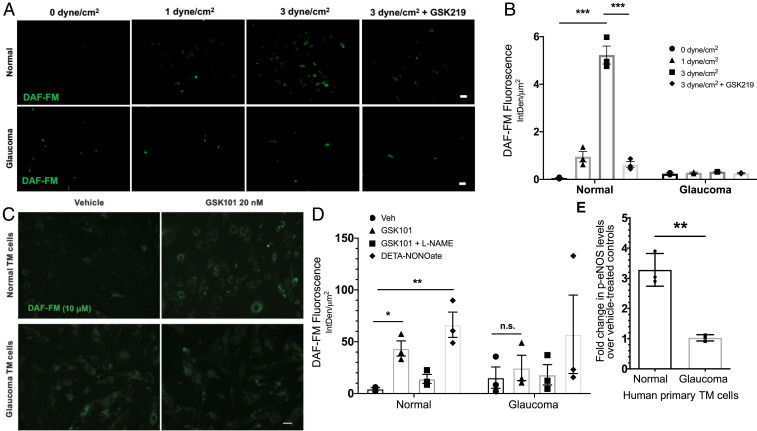
(A and B) Shear stress–mediated TRPV4-eNOS signaling is diminished in glaucomatous TM cells. (A) Normal and glaucomatous primary human TM cells were subjected to different shear stress conditions (0, 1, and 3 dyne/cm2) and treated with NO-binding DAF-FM dye to determine shear stress–mediated NO production. TRPV4 antagonist GSK219 (100 nM) was used to determine the role of TRPV4 in shear stress transduction. High shear stress (3 dyne/cm2) led to increased DAF-FM fluorescence intensity, which was reduced by TRPV4 antagonist GSK219. n = 3 normal and 2 glaucoma. (Scale bar, 50 μM.) (B) Mean DAF-FM fluorescence intensity/μm2 in normal and glaucomatous primary TM cells. ***P < 0.001, n = 3 donor strains per group; one-way ANOVA followed by Bonferroni’s post hoc test. (C–E) TRPV4-eNOS coupling is impaired in glaucomatous TM cells. (C) Representative image comparing TRPV4-mediated NO production in normal and glaucomatous primary human TM cells using DAF-FM assay. Normal and glaucomatous primary TM cells were pretreated with NO-binding DAF-FM fluorescent dye (green) and then treated with 0.001% DMSO vehicle, 20 nM GSK101, 20 nm GSK101 + 100 μM l-NAME, or 100 μM DETA/NO. n = 3/group (scale bar, 50 μm). (D) Quantification of DAF-FM fluorescence intensity/μm2 in normal and glaucomatous primary TM cells. *P < 0.05, **P < 0.01 versus the vehicle-treated group, n = 3 cell strains/group; one-way ANOVA followed by Bonferroni’s post hoc test. (E) Densitometric analysis of Western blot for p-eNOS showing relative fold change in p-eNOS levels in normal and glaucomatous primary human TM cells over their respective vehicle-treated controls. n = 3 cell strains/group; unpaired two-tailed t test. n.s., not significant.

