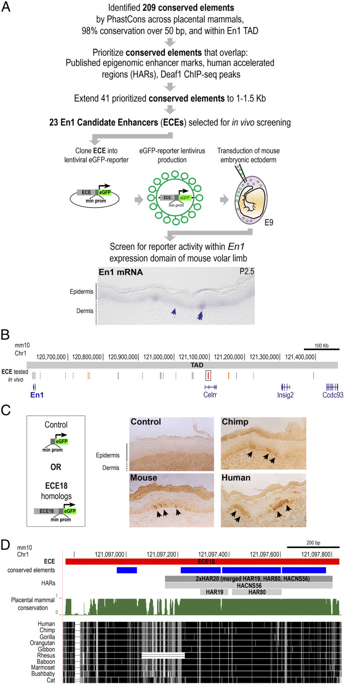
Fig. 1.
Identification of Engrailed 1 candidate enhancer ECE18. (A) Strategy to identify putative developmental En1 enhancers. Staining for En1 mRNA (purple) in mouse volar limb skin at P2.5 is shown The basal keratinocyte layer (arrow) and eccrine placode (double arrow) are shown. (B) The location in mm10 of ECEs tested in vivo (vertical gray lines). ECE18 is in red and boxed and other positive ECEs are highlighted in orange. (C) Representative images of mouse, chimpanzee, and hECE18 transgenic P2.5 volar limb stained with anti-GFP antibody. eGFP (black arrow) is visualized using HRP-DAB coupled immunohistochemistry. (D) Sequence alignment and evolutionarily features of ECE18, which contains four conserved elements by phastCons (blue rectangles) and overlaps the human accelerated regions HACNS56, HAR19, and HAR80 (collectively called 2xHAR20).

