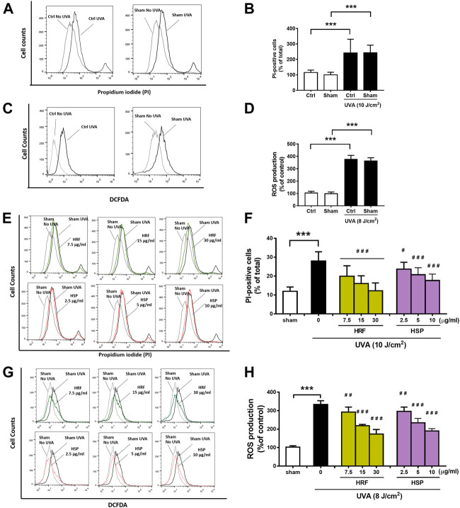
FIGURE 1.
The protective effects of HRF and its active ingredient HSP on UVA-induced cytotoxicity and oxidative stress in NHDFs. HRF at dose 7.5, 15, and 30 μg/ml and HSP at dose 2.5, 5, and 10 μg/ml as well as the ethanol treatment and no compound treatment (0), were individually treated in NHDFs prior to UVA (10 J/cm2) irradiation for cytotoxicity assay. Treated cells were harvested at 12 h post-irradiation to perform the flow cytometry of fluorescent staining, propidium iodide (PI), for the PI-positive cells (% of total). The flow histogram (A) and summary graph analysis (B) of ethanol sham treatment and compare to UVA irradiated cells and the histogram (E) and summary graph analysis of treatment groups (F). The HRF and HSP were individually treated in NHDFs prior to UVA (8 J/cm2) irradiation. Treated cells were harvested at 1 h post-irradiation to perform the flow cytometry of fluorescent staining, DCFDA dye, for the evaluation of the ROS production (% of control). The flow histogram (C) and summary graph analysis (D) of ethanol sham treatment and compare to UVA irradiated cells and the histogram were expressed as mean ± SD, n = 4. ***p < 0.001 vs. non-irradiated sham group by Student’s t-test. #p < 0.05; ##p < 0.01; ###p < 0.001 vs. the sham-irradiated group by one-way ANOVA Dunnett's test.

