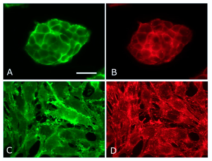
Figure 2.
Epithelial–mesenchymal transformation in NMuMg cells. Control (A,B) and syndecan-1-negative (C,D) NMuMg cells stained for β–catenin (A,C) and F-actin (B,D). Syndecan-1 was depleted by CRISPR/Cas9 technique. The loss of epithelial morphology accompanies syndecan-1 depletion, and the resulting fibroblastic morphology is accompanied by microfilament bundle formation. Scale bar = 50 µm.

